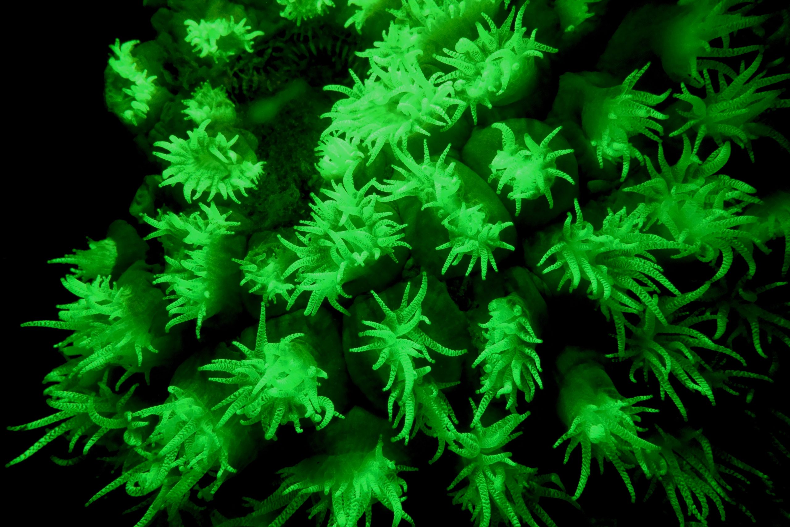
Research teams from Teva and the Technion are conducting a joint study to test the effectiveness of innovative immunotherapy treatments using antibodies or cellular therapy in unique models of cancer, Parkinson’s disease, and migraines. For example, the projects combine advanced technologies to: develop smart drug carriers that can reach the brain or cancerous tumors; activate relevant areas of activity in the brain and examine their effect on the nervous periphery and immune system, and; study the effect of the microbiome (the bacterial environment that exists within the human body) on the development of Parkinson’s disease. The projects entail a combination of antibody and protein engineering, as well as advanced computational analyses on the immune and bacterial systems.
This year, with the support of the Innovation Authority, three Nofar grants were given to researchers from the Technion that form part of the collaboration with Teva. The Innovation Authority is currently examining other similar collaborations. A total of six joint projects have been initiated in recent months by Technion and Teva researchers as part of the research collaboration.
In addition, eight outstanding students from the Technion have been selected for Teva’s national student forum known as ”The National Forum for Bio-Innovators by Teva.” The forum includes scholarships, and professional and personal training for students and their supervisors in order to connect them to the pharma world.
Managed through the Technion R&D Foundation (TRDF), these projects constitute a significant part of the cooperative program that Teva initiated and promotes with the Israeli Academy and collaborations that have been revealed during the past year. To establish these innovative collaborations, Teva conducted a thorough process to locate potential research partners and examined about 400 laboratories in the Israeli academy and about 90 startups in the field of biology. From these, Teva selected 15 strategic collaborations with leading research groups in Israeli academia. This connection creates significant and strategic value for both the academic institutions and Teva. The process is continuous and will expand over the year.
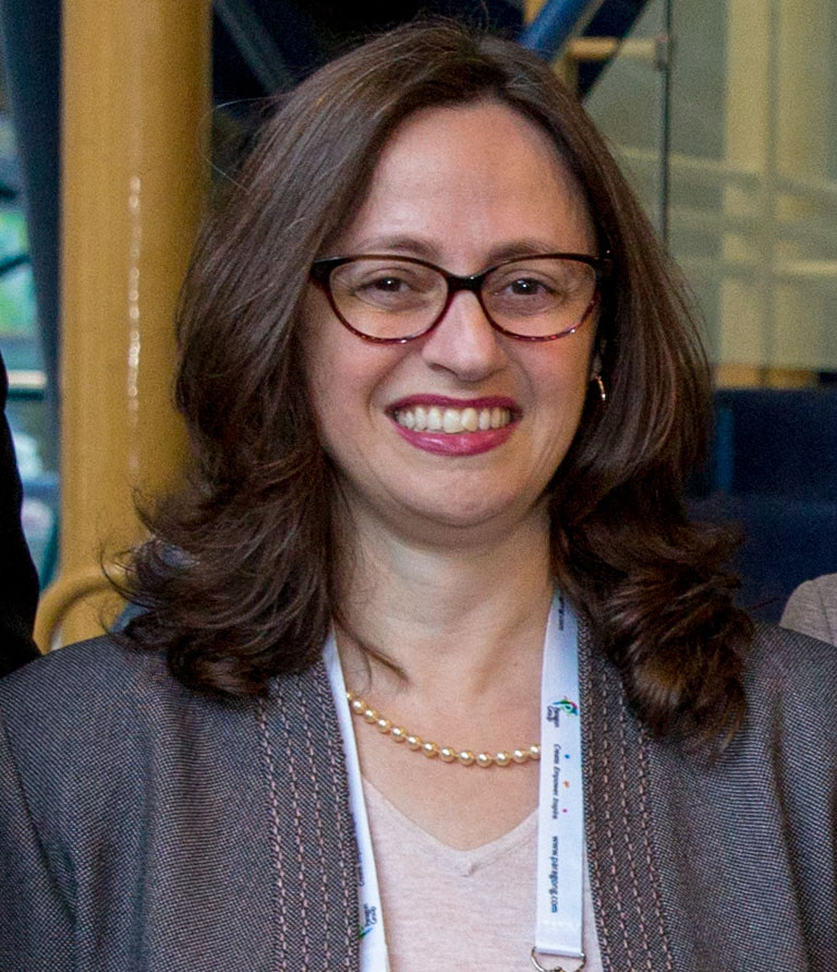
According to Dr. Dana Bar-On, project leader and director of academic relations at Teva’s Global R&D Department, ”The collaboration with the Technion is one of the key building blocks of Teva’s academic relations program – in Israel and around the world. After intensive mapping, we found leading researchers and partners in R&D at the Technion, even in the early stages. The combined force of academia and Teva is gaining momentum these days despite the delay caused by the spread of the coronavirus and the closure of some university activities. We believe the projects will advance in the coming years and some of them will be able to enter Teva’s future pipeline.”
“Collaboration between Technion researchers and Teva has borne fruit in the past and led to the development of the drug Azilect,” said Technion’s Vice President of Research Prof. Jacob (Koby) Rubinstein. ”The Technion encourages and promotes industrial and academic relations on an ongoing basis for the benefit of advancing its research and student training. I welcome the research collaboration with Teva, and I’m convinced that it will lead to innovative developments in pharma for the benefit of all mankind.”
About Teva Academia
Teva’s R&D on drug innovation began expanding in the 1980s as a result of fruitful cooperation with Israeli academia. As a result, it has developed key drugs in the company’s products portfolio that, to this day, continue to have a positive impact on the health and lives of many patients around the world.
Teva, a global company with a presence in about 60 markets worldwide, understands the strategic importance of building scientific collaborations with leading academic institutions, especially with Israeli academia. Teva believes that a close relationship with academia from the earliest stages of research can lead to the development of advanced drugs and innovative technologies for patients around the world.
Teva’s international academic activities are managed from Israel by a special team for academic affairs, led by Dr. Dana Bar-On in the company’s R&D division. Teva supports student scholarships, doctoral and post-doctoral fellowships at Teva, and joint scientific publications with academic institutions.