Predictive Medicine

Technion researchers have developed a method for predicting the safety and effectiveness of diabetes treatment methods for specific patients.  The patented discovery will help physicians determine which treatments to use and which to avoid when caring for their diabetic patients.  

Professor Andrew P. Levy, MD, Ph.D., of the Technion’s Rappaport Faculty of Medicine

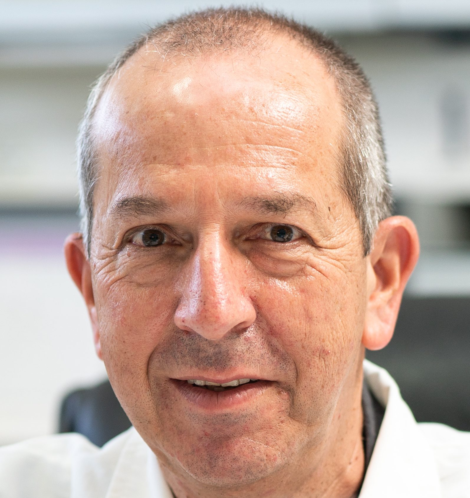
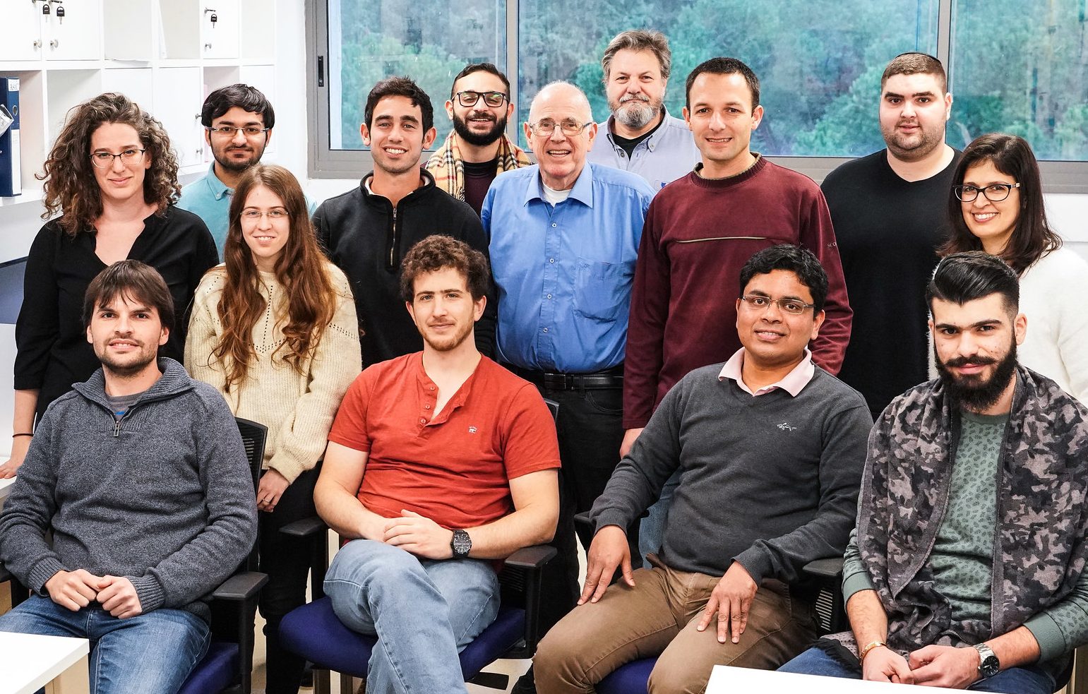
The research was led by Professor Andrew P. Levy, MD, Ph.D., of the Technion’s Rappaport Faculty of Medicine, along with Leah E. Cahill, Ph.D. and doctoral candidate Allie S. Carew of the Harvard T.H. Chan School of Public Health and Department of Medicine of Dalhousie University in Nova Scotia, Canada. The researchers investigated 5,805 diabetic patients, and their work has been patented and published in the Journal of the American College of Cardiology (JACC). 

Diabetes is one of the most common diseases in the developed world. The disease, which affects roughly 10% of Israelis, is a metabolic condition characterized by high concentrations of glucose, or simple sugars, in the patients’ bloodstream. The disease is caused by a malfunction of the insulin absorption process. When functioning correctly, the pancreas produces the hormone insulin to help cells convert glucose into energy to power the body. However, among people with diabetes, the process is disrupted for one of two reasons: either the body does not produce enough insulin, or the cells are insulin resistant. The result in both cases is hyperglycemia, an excessive accumulation of glucose in the blood.

Hyperglycemia poses a significant danger, damaging the inner blood vessel walls and leading to a narrowing of the arteries and veins. According to Prof. Levy, “Hyperglycemia harms numerous bodily functions, including vision and kidney function. However, when it impairs cardiovascular activity, the results can be catastrophic. And in fact, most deaths of diabetic patients are caused by heart disease.” 

The most common treatment of diabetes focuses on intensive therapy to reduce blood sugar levels. However, today researchers know that this drastic reduction can actually be harmful and significantly increase mortality rates. This was demonstrated a decade ago in a landmark American study called ACCORD, published by The New England Journal of Medicine.  The study showed that intensive treatment could actually increase mortality rates amongst diabetic patients. 

The revelations of the ACCORD study posed a significant ethical dilemma for doctors. Should they treat diabetes if it risks causing cardiovascular damage? 

Enter Prof. Andrew Levy, who has spent more than 20 years searching for a simple blood test for identifying diabetic patients at high risk of heart attacks. His discovery began in past research showing that certain haptoglobin protein types in diabetic patients could predict patients’ level of risk for cardiovascular complications. For example, patients with type 2-2 haptoglobin are nearly five times as likely to suffer heart attacks than patients without. Building on that data, Prof. Levy then examined the patients in the ACCORD study to see if their response to aggressive treatment correlated with their haptoglobin protein type.

The study just published in the JACC reveals that among diabetics with type 2-2 haptoglobin, an intensive reduction in blood sugar levels reduced heart attacks by approximately 30%, but did not change mortality rates. However, among diabetes patients with other types of haptoglobin protein, intensive treatment did not affect the number of heart attacks and even increased mortality rates by a dramatic 40%. 

Prof. Levy and the research team concluded that checking haptoglobin could allow doctors to predict the success of aggressive treatment. According to Prof. Levy, “The ACCORD research was curtailed because of the rise in the number of patients who died as a result of treatment. However, today we can identify the specific patients who will indeed respond positively to treatment. Currently, more studies are being planned that will verify and strengthen this research.”


Eurotech Universities Alliance

An alliance of leading European universities of science and technology is starting an international study programme with the goal of jointly shaping the engineering education of the future. This “EuroTeQ Engineering University” will be open not only to students enrolled at the partner universities, but also to engineers working in industry who are interested in life-long learning. The initiative will reinvigorate the symbiosis between society and technology together with various stakeholders and orient its programme towards human-centred engineering. The concept was now successful in the European Union’s “European Universities” Initiative.

The EU will fund the project with approximately five million euros over the next three years. The initiative emerges from the EuroTech Universities Alliance, a strategic partnership of Technical University of Denmark (DTU), École Polytechnique (L’X), Eindhoven University of Technology (TU/e), Technical University of Munich (TUM) as well as Ecole polytechnique fédérale de Lausanne (EPFL) and the Technion – Israel Institute of Technology. For this project, they have brought two other strong partners on board: Tallinn University of Technology (TalTech) and Czech Technical University in Prague (CTU). EPFL and Technion, being located in non-EU countries and hence not eligible for funding, will contribute to the implementation of the programme.

Individually designed curricula

The partners will establish a joint engineering sciences study programme across different disciplines as well as across national and institutional boundaries, reaching well beyond individual technologies. The goal of the alliance is to look at technology developments on a holistic level. “Today, we can’t talk about mobility without considering climate impacts, and robotics and artificial intelligence will not succeed without winning over human trust,” says Professor Thomas F. Hofmann, President of TUM, which coordinates the project. “A modern engineering education must provide students not only with in-depth technical knowledge but also with an extended educational horizon, an entrepreneurial mindset and socio-political sensitivity.”

In order to promote this understanding Europe-wide, the “EuroTeQ Engineering University” will not be restricted to students enrolled at the respective partner institutions. It will also be open to those who do not hold an academic degree yet, but who play an important role in value creation and communication processes. Students will be able to design their curricula in line with their learning objectives and career aspirations and benefit from new digital formats. In the spirit of life-long learning, they will be able to continue their education throughout their entire career.

Interaction between students and vocational trainees

The alliance will furthermore bring their university students together with vocational trainees pursuing a technical career and with a large number of stakeholders from industry, trade associations and various areas of society to explore the grand challenges of the 21st century and jointly formulate solution strategies. The project has already won the support of 45 collaboration partners. Thanks to this, the “EuroTeQ Engineering University” will gain comprehensive experience with the various qualification structures in Europe and will learn about the needs of the younger generation. These findings will, in turn, be integrated in the design of teaching at all partner universities.

“With EuroTeQ, we are building on the strengths of existing cooperation between the EuroTech Universities and our partners from Estonia and the Czech Republic, and on our multidisciplinary research-based curricula,” says Tatiana Panteli, Head of the EuroTech Universities Alliance Brussels Office. “We are also thrilled to add many exciting and challenging elements to this new European university, which will provide lifelong learning opportunities for students and professionals, to ‘engineer’ talents for the industry and society of the future.”

Prestigious project of the European Commission

The European Universities, introduced by French President Emmanuel Macron, are a flagship initiative of the European Commission, which aims at establishing ambitious European university alliances that will make the European university landscape even stronger in the fierce competition with the USA and Asia.

Patterns of Tentacle Movement in Coral

Researchers at the Technion and Tel Aviv University have deciphered the sophisticated movement pattern of coral tentacles, which improves the rate of food supply and oxygen fluxes around the coral. The discovery sheds light on how the species adapt to the complex reef environment.


Professor Uri Shavit of the Technion Faculty of Civil and Environmental Engineering

Researchers at the Technion and Tel Aviv University have discovered a surprising pattern to the oscillating motions of coral’s flexible appendages with respect to the wave-induced water flow in its direct vicinity. The movement pattern optimizes the coral’s oxygen and food supply. The research, published in Proceedings of The Royal Society B, was conducted by the graduate student Dror Malul and his advisors, Professor Uri Shavit of the Technion Faculty of Civil and Environmental Engineering and Professor Roi Holzman of the Department of Zoology at Tel Aviv University.

Corals are marine animals that are fixed to a substrate and therefore unable to roam in search of food. They depend on water flow for food and oxygen supply and waste disposal. Although the coral’s body is sedentary, it has tentacles that extend into the water around it, where they interact and oscillate with the waves. Corals can use their reduced muscular system and internal hydro-skeleton to extend and contract their tentacles to catch prey or defend against predators but are unable to move the tentacles from side to side.

Professor Roi Holzman of the Department of Zoology at Tel Aviv University.

The researchers filmed the coral tentacle movement underwater, at the coral reef, and in a laboratory wave facility at the Interuniversity Institute for Marine Sciences in Eilat, Israel. They made an unexpected discovery that the oscillating movements of the tentacles are not passive. In other words, the coral tentacles precede the movement of the waves in an out-of-phase motion.

According to Prof. Shavit: “In the experiments, we measured the movement of the tentacles and the speed of water near them, and we were surprised to find that the tentacles precede the motion of the water. They move at the same frequency as the wave frequency, but with a phase difference of nearly ¼ of the wave cycle. The result was intriguing because it was clear to us that corals do not have a muscular system capable of moving the tentacles sideways.”

The study shows that the out-of-phase motion results from the tentacles’ elasticity, which can presumably be modified by the animal. At the beginning of the motion, the elastic tentacles move with the wave. Towards halfway of the cycle, the water flow weakens and the elastic force returns the tentacle to its central position before the water speed changes its direction again. Using mathematical modeling, simulations, and other measurements, the researchers found that the phase difference and the movement of the tentacles relative to the water improve three important functions: the amount of oxygen the coral absorbs at night; removal of excess oxygen during daily photosynthesis; and the rate of food supply (plankton).

Dror Malul

Photographing the tentacle movement of other coral species suggests that this mechanism is typical of all coral tentacles. According to Dror Malul, “In the dense and competitive environment of the reef, any mechanism that provides the coral with a competitive advantage over its neighbors can determine which of the coral species will survive. To date, many biological mechanisms that help coral adapt to the complex reef environment have been revealed, but the path to fully understanding all of its physical mechanisms is still long.”

Following the discovery, the researchers focused on the following areas: learning the mechanism that creates the phase difference by developing a dynamic model for calculating the periodic movement of coral tentacles; developing a method of measuring the elastic properties of the tentacles, and; exploring the mechanisms that influence the ability of corals to increase the inlet and outlet fluxes depending on water velocity and the phase difference. The researchers postulate that the out-of-phase motion and the resulting elevated fluxes are general and occur not only with coral tentacles but also in river vegetation, natural forests, and agricultural fields.

Click here for the paper in Proceedings of the Royal Society 

Accelerating Blood Test Analysis 98%

Professor Tomer Shlomi

Researchers at the Technion have developed a new method for rapid and inexpensive analysis of the chemical composition of blood samples, which may hasten the early diagnosis of diseases. The first application to be tested will be the early detection of various cancerous tumors based on blood tests.

The innovative technology, which was published in Nature Communications, was developed by Professor Tomer Shlomi and doctoral students Shoval Lagziel and Boris Sarvin. It is based on a unique combination of mass spectrometry and computational methods developed by the research group.

A mass spectrometer is a common device used to determine the concentrations of molecules in biological samples. Testing using this device typically requires a preliminary process called chromatography that entails the separation of the materials in the sample according to chemical properties.

Doctoral student Shoval Lagziel

Chromatography, which increases the sensitivity of the spectrometric measurement, is time-consuming and therefore makes the process expensive. One sample typically costs hundreds of dollars. As a result, it is desirable to find a way to skip the chromatographic step without compromising the sensitivity of the analysis, that is, the ability to identify many molecules and quantify their concentrations.

In the current study, Prof. Shlomi’s research group presents a method that skips the chromatography step and makes it possible to directly use mass spectrometry without significantly impairing the quality of the analysis. The test is completed in just 30 seconds, thus shortening the process by about 98% and reducing its cost by a similar rate. 

According to Prof. Shlomi, the novelty lies in the use of a computational method developed by the research group. They employ a method that identifies optimal working configurations in the mass spectrometer, which allows for a high-sensitivity analysis for specific types of biological samples.

Doctoral student Boris Sarvin

The computational analysis also corrects the measured raw information and accurately quantifies concentrations of thousands of molecules in blood samples.

Prof. Tomer Shlomi is a faculty member in the Faculties of Computer Science and Biology and a member of the Lorry I. Lokey Center for Life Sciences and Engineering. The research was funded by an ERC grant and by the Israel Science Foundation.

For the full article on Nature Communications click here.

BOARD OF GOVERNORS 2020

Vision 2020 – Meet brilliant Technion researchers in their labs.

Come take part in the Technion International Board of Governors for 2020. This is the year of new vision, new possibilities, and new dimensions of engagement.

Tune in live with the Technion global family!

LIVE NOW:

https://www.facebook.com/Technion.Israel/videos/3300368766855358/

BOG 2020


Ion-specific membranes

Inspired by the potassium channel in cells: The future of ion-selective synthetic membranes is vast

A Nature Nanotechnology perspective piece highlights the importance of ion-specific membranes and “paves pathways for new research avenues.”

Professor Menachem Elimelech (on the right) and Assistant Professor Razi Epsztein

A Nature Nanotechnology perspective piece by new Assistant Professor Razi Epsztein, of the Technion Faculty of Civil and Environmental Engineering, and his postdoc mentor, Professor Menachem Elimelech from Yale University, highlights the future of synthetic membranes, which are used widely in desalination and other technologies. The focus of the researchers’ piece is on the future fabrication of membranes with sub-nanoscale pores that possess the ability to selectively distinguish between specific ions in water, even extremely similar ones, such as sodium and potassium.

Currently, membrane technology is predominantly used in reverse osmosis desalination, an energy-efficient way to ideally remove all salts from water. However, reverse osmosis and other types of synthetic membranes lack the ability to discriminate between ions, which results, for example, in the redundant step of re-adding the ions that are necessary for drinking water to be safe. Moreover, the waste, or brine, resulting from current non-selective desalination is an environmental concern.

Based on the potassium channel found in cells, synthetic ion-selective membranes take their inspiration from nature’s ingenuity. Such ion-selective technology would pave the way for membranes that can be used to remove a contaminant from groundwater while leaving “good” ions behind; membranes with the ability to mine important elements from water such as lithium, which is a valuable resource for batteries in our wireless world; a method to pre-treat and remove calcium and magnesium from seawater, before desalination, to reduce scale buildup that shortens the lifespan of reverse osmosis membranes; filtering or sensing target compounds in medicine; and much more.

Emphasizing the potential of ion-selective membranes, Prof Epszstein said that, “Selectivity is fascinating and important at the same time. Improving our ability to discriminate and separate between small ions and molecules can be super beneficial for water-treatment processes, as well as for resource recovery, energy production, sensing, and even medicine.”

Prof. Epsztein’s lab will focus on the development of “selective separation technologies for a wide range of environmental applications, with a focus on membrane technologies at the water-energy nexus.” This will entail the fundamental study of transport in polymeric (organic) membranes and advanced materials followed by the fabrication of selective membranes for various applications that require high selectivity.

Photosynthesis for Clean Energy

In a big step forward toward making BIO-photoelectrochemical cells (BIOcells) a mainstream clean energy source in the future, researchers from the Technion – Israel Institute of Technology and Ruhr-Universität Bochum (RUB) have overcome an efficiency hurdle by successfully combining the power of efficient light absorption by photosynthetic light-harvesting complexes with the electrochemical power of Photosystem II (PSII), nature’s water splitting enzyme. The breakthrough is a functional solution to overcoming previously limited efficiency due to a “green-light gap” in the absorption spectrum of biosolar energy devices. The findings were published in the Journal of Materials Chemistry A.

Tapping into the power of photosynthesis for clean energy

As the world strives to replace fossil fuel with clean energy sources, solar energy – because of its abundance and total lack of polluting elements – is considered a particularly valuable energy source. In nature, bacteria, algae, and plant life have evolved to efficiently convert solar energy into chemical energy via photosynthesis. BIOcells are an innovative concept in the field of renewable energy aimed at harnessing this natural process semi-artificially for the development of clean, affordable, and efficient energy sources.

Professor Noam Adir

BIOcells utilize large protein complexes called photosystems, which have the capacity to convert sunlight into electrical energy. Isolated from plants, algae, or cyanobacteria, photosystems are responsible for natural sunlight to energy conversion in nature. PSII is a valuable type of photosystem because it uses water as an electron source for the generation of electricity. It is the source of all the oxygen that we breathe and all the food that we eat. 

But BIOcells containing only PSII complexes only have limited efficiency. The efficiency is measured by the amount of electrical power coming out of the cell divided by the sunlight energy coming in, and PSII alone can only convert a limited range of light. They are unable to convert green light, which constitutes about 50% of visible light, into energy. In cyanobacteria and red algae, this is rectified by the Phycobilisome (PBSs) light harvesting complex. PBSs are protein structures found in cyanobacteria that enable them to harvest light that is not absorbed efficiently by the chlorophyll molecules in PSII. PBSs function as a light-absorbing transmitter, directing excitation energy into the reaction centers of PSII.

“As unique as PSII is, its efficiency is limited, because it can use merely a percentage of the sunlight,” explained Professor Marc Nowaczyk, head of the Molecular Mechanisms of Photosynthesis project group at RUB. “Cyanobacteria have solved the problem by forming special light-collecting proteins, i.e. the PBS, which also make use of this light. This cooperation works in nature, but not yet in the test tube.” Professor Noam Adir of the Schulich Faculty of Chemistry added that, “just as in nature, our two groups collaborated, bringing our expertise in isolating the PBS with Prof. Nowaczyk’s groups expertise in isolating PSII. Together we overcame the obstacles of putting it all together in the BIOcell.”

In order to make the collaboration between cyanobacteria and plant photosynthesis functional in an artificial BIOcell, the two teams succeeded in producing a two-component bioelectrode. This included the difficult task of functionally joining the PBS and PSII multiprotein complexes.  some of which were combined across species. 

The researchers, led by Dr. Volker Hartmann (RUB) and Dr. Dvir Harris (Technion), stabilized the interaction between PSII and PBS by permanently fixing the proteins at a very short distance from each other using crosslinkers. Crosslinkers are molecules with two or more reactive ends that are capable of chemically attaching to specific functional groups on proteins. After crosslinking PSIIs with PBSs, the team was then able to insert the super complexes into the appropriate electrode structures. 

Dr. Dvir Harris

Integration of the PBS–PSII super-complexes within a hydrogel on macro-porous indium tin oxide electrodes (MP-ITO) improved the incident photon-to-electron conversion efficiencies (IPCE). IPCE values in the “green gap” were doubled compared to PSII electrodes without PBS and the IPCE in the green light gap reached a maximum of 10.9%. 

The capacity to assemble these proteins is a breakthrough in biological solar cell development. This means that protein complexes from different species can be functionally combined to create semi-artificial systems that have the cumulative advantages of the different species utilized. 

In their future BIOcell research, the teams will mainly focus on optimizing the production and life span of the biological components. 

The research was funded by the German-Israeli research project Nano-engineered Opto-bioelectronics with Biomaterials and Bio-inspired Assemblies under the auspices of the German Research Foundation (DFG) and the Israel Science Foundation and the Ruhr Explores Solvation Resolv Cluster of Excellence (www.solvation.de) and the GRK 2341 Microbial Substrate Conversion Research School (Micon), which is financed by the DFG.

Professor Noam Adir is a member of The Nancy and Stephen Grand Technion Energy Program (GTEP), and Dr. Dvir Harris is a GTEP PhD track graduate.

Click here for the paper in Journal of Materials Chemistry A

Cancer & the Heart

There is a connection between heart disease and cancer, a novel study has revealed. The findings, published in Circulation, could potentially help cardio-oncologists slow cancer progression and improve cancer outcomes. Researchers led by Professor Ami Aronheim, Professor Yuval Shaked, and Dr. Shimrit Avraham have determined that early changes in the heart resulting from cardiac disease or damage (cardiac remodeling) promotes cancer progression. 

Professor Ami Aronheim and Dr. Shimrit Avraham

Recently, studies have indicated that cancer and cardiovascular diseases are connected, and that heart failure and stress correlate with a poor cancer prognosis. While it has been determined that chemotherapy drugs can be damaging to heart muscle, the effects of cardiac remodeling on cancer are not well known. To uncover the connection between cardiac remodeling and cancer, a research team led by Professor Ami Aronheim, Professor Yuval Shaked, and Dr. Shimrit Avraham, from the Rappaport Faculty of Medicine at the Technion, and their colleagues Professor Walid Saliba and Professor Avinoam Shiran from Carmel Hospital, have investigated whether early cardiac remodeling in the absence of heart failure promotes cancer. 

Professor Ami Aronheim,

To mimic cardiac remodeling, the research team collaborated with the Preclinical Research Authority, led by Dr. Rona Shofti and Dr. Tali Haas, and used a laboratory technique called transverse aortic constriction (TAC) to exert mechanical pressure on the hearts of laboratory mice. TAC stresses the mouse heart with a pressure overload resulting in an increase in heart cell growth called hypertrophy, which is a common effect of cardiovascular complications. The team then implanted cancer cells into the TAC-operated mice to see if the early cardiac remodeling affects tumor progression.

The researchers found that the TAC-operated mice developed larger tumors at the site of the implanted cancer cells. In addition, TAC-operated mice displayed a higher rate of cancer cells seeding to the lungs, representing metastases (secondary tumors spread from the original lesion) as compared to non-operated mice. 

The investigators also found that serum from TAC-operated mice resulted in enhanced cancer cell proliferation in cell cultures (in vitro), suggesting that tumor-promoting proteins are present in the blood from the TAC-operated mice. Specifically, a protein called Periostin, which is highly expressed in the hearts of the TAC-operated mice. To investigate the effects of Periostin, on cancer cells, the researchers studied how it affected cancer cells in vitro. They found that the addition of purified Periostin enhanced cancer cell proliferation, and that the depletion of Periostin from mouse serum lowered cancer cell proliferation (in vitro). 

Dr. Shimrit Avraham

The results of the study, published in Circulation highlight the connection between cardiovascular disease and cancer, and the importance of early diagnosis and treatment of cardiac disease in cancer patients. Such intervention has the potential to significantly attenuate cancer progression and improve cancer outcomes. 

“As a result of the study, we recommend that you treat heart problems early, when the body is still successfully coping with the problem, and not wait for a chronic condition,” said Prof. Aronheim. “Such problems can be detected with a simple echocardiography test, and in many cases, early catheterization may help to slow cancerous development.”

The research was supported by the Israel Science Foundation (ISF). 

Click here for the paper in Circulation  


Computational Computer Memory

The study is a significant milestone towards the development of computer memory units with computational abilities, a solution to the call for faster processing and analytics needed for Big Data.

Researchers at the Viterbi Faculty of Electrical Engineering have presented a breakthrough in in-memory computing: the use of computer memory to perform computational operations. The study, published in IEEE Transactions on Electron Devices, was led by Professor Shahar Kvatinsky, and graduate student Barak Hoffer, in collaboration with Professor Rainer Waser’s group from the Jülich Research Center and researchers Dr. Vikas Rana and Dr. Stephan Menzel.

Prof. Shahar Kvatinsky Research group

Since the first computers were built in the 1940s, the basic structure of the computer has hardly changed.

“The classic computer we are familiar with consists of two central units — the processor, which performs computations and the memory, which stores the information,” said Prof. Kvatinsky. “Over the past decades, in both of these areas, meteoric improvements have been achieved – the processing ability of the processors has increased significantly and the storage capacity in the memory units has increased dramatically. But communication between them has become a bottleneck that limits the entire computer’s processing rate. This is because the transfer of information between the processor and memory is significantly slower than the processing itself and consumes a lot of energy. ” 

In-memory computing is based on performing digital computing operations, similar to those done by the processor, by the memory units themselves, and not in a dedicated processing unit. This may lead to substantially faster performance as compared to the classic computer.  As the amount of data increases, and the demand for faster processing and analytics on big data grows, in-memory computing is a desirable solution: By both storing data and processing it in parallel, in-memory computing allows businesses to deliver immediate action.

In recent years, Prof. Kvatinsky’s group has focused on several avenues to achieve functional in-memory computing. The memristor-aided logic (MAGIC) technique, invented by Prof. Kvatinsky, does this by employing memristive devices – a form of circuit that can both store and process information and exceeds the performance of conventional circuit technologies in this domain. The present article demonstrates how in-memory computing can be achieved using the MAGIC technique on fabricated memristor devices.  

“The attempt to process data in the memory unit is not entirely new,” explained Prof. Kvatinsky, “but its technological realization is very complex and challenging, partly because of the different physical properties of the computational components in the processor (transistors) and the components exist in computer memory today. Some attempts have been based on performing some of the computation close to the storage units, thus reducing the cost of transferring information between the units. Attempts have also been made to use the memory cells for computation, but this is usually a limited calculation that differs in essence from the digital calculation performed by the processor. “

In the article, Technion researchers present a successful assimilation of three logical gates within a memory unit created by the Jülich partners, and demonstrate, experimentally, that “the new gates produce correct and reproducible results.” In addition, they demonstrate the feasibility of more complex logic functions. Prof. Kvatinsky believes this is a significant milestone towards the development of computer memory units with significant computational abilities. 

“For about a decade we’ve been developing theory and computer simulations of how the logic gates we designed will perform logical calculations in a similar way to a processor,” he said. “This is the first work that demonstrates this method on memory components created and measured in the lab, moving the method from theory to practice.”

The study was conducted with the support of the European Research Council (ERC Starting Grant) and the Israel Science Foundation.

For the full article in IEEE Transactions on Electron Devices click here


See the Light

The beautiful phenomenon allows for new and exciting research opportunities in the field of Optics and Optofluidics


Haifa, Israel July 2, 2020 – A team of researchers from Technion – Israel Institute of Technology has observed branched flow of light for the very first time. The findings are published in the prestigious scientific journal Nature and are presented on the cover of the July 2, 2020 issue.

Distinguished Professor Mordechai (Moti) Segev

The study was carried out by Ph.D. student Anatoly (Tolik) Patsyk in collaboration with Miguel A. Bandres, who was a postdoctoral fellow at Technion when the project started and is now Assistant Professor at CREOL, College of Optics and Photonics, University of Central Florida. The research was led by Technion President Prof. Uri Sivan and Distinguished Prof. Mordechai (Moti) Segev of Technion’s Physics and Electrical Engineering Faculties, the Solid State Institute, and the Russell Berrie Nanotechnology Institute.  

When waves travel through landscapes that contain disturbances, they naturally scatter, often in all directions. Scattering of light is a natural phenomenon, found in many places in nature. For example, the scattering of light is the reason for the blue color of the sky. As it turns out, when the length over which disturbances vary is much larger than the wavelength, the wave scatters in an unusual fashion: it forms channels (branches) of enhanced intensity that continue to divide or branch out, as the wave propagates.  This phenomenon is known as branched flow. It was first observed in 2001 in electrons and had been suggested to be ubiquitous and occur also for all waves in nature, for example – sound waves and even ocean waves. Now, Technion researchers are bringing branched flow to the domain of light: they have made an experimental observation of the branched flow of light.

Technion President Professor Uri Sivan

“We always had the intention of finding something new, and we were eager to find it. It was not what we started looking for, but we kept looking and we found something far better,” says Asst. Prof. Miguel Bandres. “We are familiar with the fact that waves spread when they propagate in a homogeneous medium. But for other kinds of mediums, waves can behave in very different ways. When we have a disordered medium where the variations are not random but smooth, like a landscape of mountains and valleys, the waves will propagate in a peculiar way. They will form channels that keep dividing as the wave propagates, forming a beautiful pattern resembling the branches of a tree.” 

In their research, the team coupled a laser beam to a soap membrane, which contains random variations in membrane thickness. They discovered that when light propagates within the soap film, rather than being scattered, the light forms elongated branches, creating the branched flow phenomenon for light.

Observation of branched flow of light

“In optics we usually work hard to make light stay focused and propagate as a collimated beam, but here the surprise is that the random structure of the soap film naturally caused the light to stay focused. It is another one of nature’s surprises,” says Tolik Patsyk. 

The ability to create branched flow in the field of optics offers new and exciting opportunities for investigating and understanding this universal wave phenomenon.

Anatoly Patsyk

“There is nothing more exciting than discovering something new and this is the first demonstration of this phenomenon with light waves,” says Technion President Prof. Uri Sivan. “This goes to show that intriguing phenomena can also be observed in simple systems and one just has to be perceptive enough to uncover them. As such, bringing together and combining the views of researchers from different backgrounds and disciplines has led to some truly interesting insights.”

“The fact that we observe it with light waves opens remarkable new possibilities for research, starting with the fact that we can characterize the medium in which light propagates to very high precision and the fact that we can also follow those branches accurately and study their properties,” he adds. 

Thin liquid membranes as a platform for observing branched flow of light

Distinguished Prof. Moti Segev looks to the future. “I always educate my team to think beyond the horizon,” he says, “to think about something new, and at the same time – look at the experimental facts as they are, rather than try to adapt the experiments to meet some expected behavior. Here, Tolik was trying to measure something completely different and was surprised to see these light branches which he could not initially explain. He asked Miguel to join in the

Assistant Professor Miguel Bandres Photos: Nitzan Zohar, Technion spokesperson's office
Assistant Professor Miguel Bandres
Photos: Nitzan Zohar, Technion spokesperson’s office

experiments, and together they upgraded the experiments considerably – to the level they could isolate the physics involved. That is when we started to understand what we see. It took more than a year until we understood that what we have is the strange phenomenon of “branched flow”, which at the time was never considered in the context of light waves. Now, with this observation – we can think of a plethora of new ideas. For example, using these light branches to control the fluidic flow in liquid, or to combine the soap with fluorescent material and cause the branches to become little lasers. Or to use the soap membranes as a platform for exploring fundamentals of waves, such as the transitions from ordinary scattering which is always diffusive, to branched flow, and subsequently to Anderson localization. There are many ways to continue this pioneering study. As we did many times in the past, we would like to boldly go where no one has gone before.” 

The project is now continuing in the laboratories of Profs. Segev and Sivan at Technion, and in parallel in the newly established lab of Prof. Miguel Bandres at UCF. 


Click here for the paper in Nature

Click here for video demonstrating the research

Food Innovations

International Technion Innovation: Technologies to reduce sugar consumption, create healthy plant-based alternatives to unhealthy, animal-based stabilizers, and prevent food poisoning

The EU has allocated millions of euros to three multinational research teams that include researchers from the Technion Faculties of Biotechnology and Food Engineering and Mechanical Engineering. These researchers are participating in EIT-FOOD projects with cumulative budgets of more than 2.25 million, supported by grants of the EU.

The objective: To promote inventions that improve food quality and human health.

Three teams involving Technion – Israel Institute of Technology researchers are running projects of around € 1 million each supported by EU grants from the EIT-Food consortium,  the leading food innovation initiative of the European Union whose goal is to lead a revolution in food innovation, business creation, and education.

Sugar-Out, Prote-In

Professor Yoav D. Livney

A consortium led by Professor Yoav D. Livney, of the Technion Faculty of Biotechnology and Food Engineering, is fighting diabetes and obesity by developing the first healthy sweetener for the food & beverage market. The healthy, zero-glycemic-index protein-based sugar-substitute has the potential to revolutionize the global food and beverage market.

The consortium also includes PepsiCo, Danone, and Amai Proteins, a startup led by Dr. Ilan Samish. Amai Proteins is a member of the “Rising Food Stars” startup club of the EIT-FOOD.

 “The EIT Food, which the Technion is a partner of, is revolutionizing the European food ecosystem. Our project within this consortium is expected to bring to the global market an innovative sweet protein, along with a novel microencapsulation technology, to replace sugar, a major cause of obesity and diabetes (which are also risk factors for COVID-19 mortality). Sugar replacement is a tough challenge, and there is a great need for non-artificial intensive sweeteners, with a sensory profile similar to that of sugar, that is suitable for the huge global food & beverage market,” said Prof. Livney.

High-pressure processing to achieve a healthy plant-based alternative to unhealthy stabilizers 

Assistant Professor Avi Shpigelman

The Laboratory for Novel Food and Bioprocessing, led by Assistant Professor Avi Shpigelman, is partnering with the EIT project “HPHC – Development and application of hydrocolloids functionalized by dynamic high pressure.” The project aims to create healthier nutrition by physically modifying common currently used polysaccharide-based hydrocolloids using high-pressure food processing to achieve an improved range of techno-functionalities. The goal is to replace or reduce currently used additives and stabilizers with plant-based materials. 

The technology is based on ultra-high-pressure homogenization (UHPH), where a liquid is continuously pumped through a narrow valve using high pressures of up to 350 MPa. This results in the modification of biopolymer structure. This technology was originally developed for the pasteurization and sterilization of liquid foods. Specifically, the project aims to physically modify plant-based polysaccharides and fibers with the intention to replace animal-based or unhealthy stabilizers with plant-based, health-promoting ingredients. The German Institute for Food Technology (DIL) is leading the initiative together with the Technion, Herbstreith & Fox, Maspex, ZPOW Agros Nova, and Glucanova.

According to Dr. Spiegelman, “We believe that the project will increase the assimilation of diverse hydrocolloids from plant sources into the food industry and will expand the use of these materials for a healthier diet for the population. “

Lab on a chip – an early warning platform for food safety

Professor Yechezkel Kashi

Professor Yechezkel Kashi of the Faculty of Biotechnology and Food Engineering and Professor Gilad Yossifon of the Faculty of Mechanical Engineering are leading this consortium with six European partners (EUFIC, Grupo AN, Maspex, Energy Pulse Systems, and the University of Queen Belfast) to develop a technology to improve food safety by rapid monitoring of pathogenic bacteria and toxins.

Food poisoning leads to thousands of deaths each year. Food contaminants are mostly monitored in plants using culture-based and time-consuming methods of up to one week. By that time, some of the contaminated products are released to the market and consumed. The Technion team has developed a technology for sensitive and real-time detection of different pathogens and toxins, based on the “lab-on-a-chip” technology developed and verified by the Technion. This technology includes the concentration of bacteria and amplification of their DNA sequences until a measurable signal is obtained. 

Professor Gilad Yossifon

“Our goal is to integrate the technology into food product tests to obtain safety evaluation in real-time,” said Prof. Kashi. “Our solutions will improve societal well-being, minimize recalls and food outbreaks, and improve production efficiency. It is also relatively inexpensive compared to the existing methods, so it will encourage the parties involved in the food market to check products frequently throughout the supply chain, eliminating contaminated raw materials and products in real-time. This will help to avoid recall events, which are harmful for companies and their images.”


Autonomous Microscope

Technion researchers have developed an innovative microscopic method based on deep learning and self-design of the optical system, enabling the study of dynamic 3D images at super-resolution. The ability to study the dynamics in whole cells over such times scales was rarely possible until now.   


Researchers at Technion – Israel Institute of Technology present a breakthrough in 3D super-resolution microscopy of cells in the journal Nature Methods. The innovative system significantly shortens 3D image acquisition time by using a neural network and deep learning. The researchers experimentally demonstrated system efficiency in 3D mapping of mitochondria (the cell’s energy maker) and volumetric imaging of fluorescently labeled telomeres (chromosome edge regions, which are responsible, among other things, for cell division in the body) in live cells.

The research was carried out by Asst. Prof. Yoav Shechtman and Ph.D. student Elias Nehme, of the Faculty of Biomedical Engineering together with Asst. Prof. Tomer Michaeli of the Viterbi Faculty of Electrical Engineering.

Asst. Prof. Yoav Shechtman

A major challenge in biology today is the super-resolution mapping of dynamic biological processes in living cells. That is, mapping with a resolution 10 times greater than that of a standard optical microscope.

Microscopes, as a rule, produce two-dimensional images. Information is innately missing from such images as the world is three-dimensional. Currently, 3D images are usually obtained through layer scanning – the imaging of different layers in the sample and their computerized integration into a 3D image. This process is problematic as it requires a long scanning time, during which the object being examined must be static. In addition, in classical optical microscopy, the level of resolution (separation capacity) is limited by the “diffraction limit” formulated by German physicist Ernst Karl Abbe in 1873.

Enter DeepSTORM3D – a super-resolution 3D mapping system developed by the researchers. According to Asst. Prof. Yoav Shechtman, who led the development of DeepSTORM3D, “To get depth information from a 2D image we use wavefront shaping – an optical method that encodes the depth of each molecule in the image obtained on the camera. The problem with this method is that if several molecules are close by, their images overlap on the camera, and this drastically impairs spatial and temporal resolution, to the point that some samples cannot produce useful images at all.”

Asst. Prof. Tomer Michaeli

To address this challenge, researchers harnessed the field of deep learning and developed an artificial neural network – a system that performs computational tasks at unprecedented performance and speed. Together with Asst. Prof. Tomer Michaeli, an expert in this field, the researchers developed a neural network able to generate and train itself using a large number of virtual samples, and then produce super-resolution 3D images from microscopy data of real samples.

According to Shechtman, “The new technology has advanced us towards realizing one of the holy grails of biological research – mapping biological processes in living cells in super-resolution. It is important that the life sciences benefit from our instrumentation, and we maintain close relationships with biologists who explain their needs to us.”

Shechtman used the neural networks not only to analyze the images but to also improve the instrumentation. “This is perhaps the most exciting direction to emerge from the current development – the neural network has provided us with the optimal physical design of the optical system. In other words – the computer not only analyzes the data but has shown us how to build the microscope. This concept can also be applied in non-microscopy-related fields, and we are working on it.” 

Ph.D. student Elias Nehme

Research participants include Dr. Daniel Freedman from Google Research, and researchers and students from the Faculty of Biomedical Engineering, the Lorry I. Lokey Interdisciplinary Center for Life Sciences and Engineering, and the Russell Berrie Nanotechnology Institute: Racheli Gordon, Boris Ferdman, Dr. Lucien E. Weiss, Dr. Onit Alalouf, Tal Naor, and Reut Orange. The research was conducted with the support of the European Research Council Horizon 2020 Program, Google, the Israel Science Foundation and the Zuckerman Foundation. Asst. Prof. Yoav Shechtman is a Zuckerman Faculty Scholar and Dr. Lucien E. Weiss is a Zuckerman Postdoctoral Fellow.

Click here for the paper in Nature Methods