


Invisible Particles That Control Star Birth Measured for First Time
Technion-led team achieves first measurement of cosmic rays deep inside star-forming nebula 400 light-years from Earth

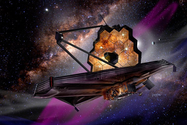
Technion-led team achieves first measurement of cosmic rays deep inside star-forming nebula 400 light-years from Earth

Symposium: When Science Meets the Field
10.02.2026 Tuesday, at 08:30
Add to calendar

Technion on the Bar - When Waste Meets Recycling
17.02.2026 Tuesday, at 20:00
Add to calendar

The Second Israeli Conference on the Philosophy of Artificial Intelligence
04.03.2026 Wednesday, at 09:30
Add to calendar
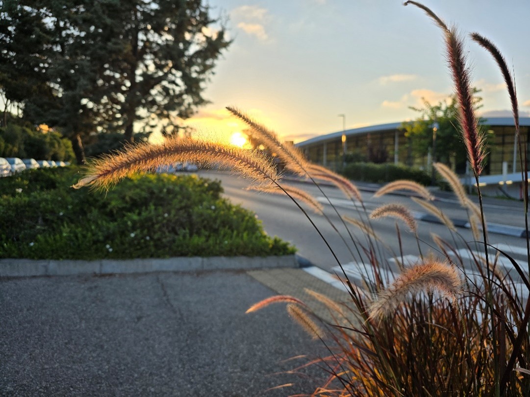
Nature on Campus Photography Exhibition
04.01.2026 Sunday, at 09:00
Add to calendar

Landmarks Exhibition
01.01.2026 Thursday, at 09:00
Add to calendar
15000
Students
in a variety of degrees
60
Research Centers
across campus
18
Faculties
100000
Graduates