
Between the Sexes
Researchers in the Technion Faculty of Biology Discover a Sexual Dimorphism in the Structure of a Single Neuron, with a Role in Sexual Behavior in C. elegans
Is there a difference in brain structure between men and women? And if we were to find such a difference in a single neuron, would it matter?
One of the most useful models for studying these questions is the nematode Caenorhabditis elegans (C. elegans). This tiny worm has several characteristics that make it an excellent research model, one of which is that every cell in its body has a predetermined identity and lineage. Like humans, C. elegans has two sexes. However, instead of male and female, the two sexes of this worm are male and hermaphrodite—a self-fertilizing individual capable of producing both male and female gametes (sperm and eggs), allowing it to reproduce without a partner.
Researchers from the Faculty of Biology at the Technion-Israel Institute of Technology examined these sex-specific differences (sexual dimorphism) in C. elegans, and uncovered surprising findings. The study, published in Proceedings of the National Academy of Sciences (PNAS), was led by Dr. Yael Iosilevskii and Dr. Menachem Katz from Prof. Beni Podbilewicz’s Lab, in collaboration with Prof. David H. Hall of the Albert Einstein College of Medicine in New York.

The researchers discovered that a highly branched neuron called PVD, previously characterized in hermaphrodites, forms a different structure in males. Moreover, while in hermaphrodites, PVD functions primarily in pain sensing, in males, it has an additional role during mating; when its development is disrupted, males are slower and less coordinated. This discovery provides a unique example of sexual dimorphism in the structure of a single neuron, which is linked to behavioral differences.
“Male” vs. “Female” Brains
It has long been established that men and women have different susceptibilities to various neurological disorders. For example, women are more prone to depression, while men have a higher risk of Parkinson’s disease. Could these differences be linked to the structure of individual neurons in the brain? This is difficult to determine due to the sheer number of neurons in the human brain – approximately 75 billion (von Bartheld et al., 2016). Even if a difference were found between the sexes in just one neuron, pinpointing its exact contribution would be challenging, as even the simplest tasks require a multitude of intricately interconnected neurons.
To explore the significance of a single neuron’s spatial structure, researchers have turned to the nematode C. elegans, just one millimeter long. A unique feature of this organism is that the identity of all 302 neurons in the hermaphrodite is invariant, allowing scientists to map their placement, spatial structure, and connections fully. “Furthermore,” said Prof. Podbilewicz, “within the nematode population, there are also male individuals with distinct anatomy, additional neurons, and different behavior. This makes for a remarkably simple system where we can directly ask: What determines the structure of each neuron in the nervous system? Are there sex-specific differences, and do they affect behavior?”
To answer these questions, Dr. Iosilevskii and Dr. Katz studied the development of the sensory neuron PVD. This neuron has a highly branched structure, with repetitive subunits resembling a candelabra (“menorahs”). Its distinctive shape and its development during the organism’s maturation have made it a research focus for over a decade. While much is known about its development in hermaphrodites, PVD had not been characterized in males or examined for sexual dimorphism. The Technion researchers set out to determine whether male PVD neurons develop a different spatial structure and whether this difference influences a male’s behavior.
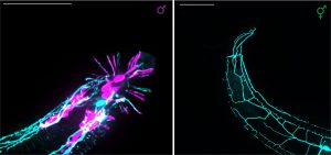
When examining PVD development in males, the researchers found that its menorah-like structures remained consistent across both sexes. However, they were surprised to discover that in adult males, PVD extends additional branches into the tail fan – a specialized male organ used for mating. Along with Prof. Hall, they found that these branches are entirely separate from the previously known neurons in this region. This unique branching of PVD does not occur during the tail fan’s development but emerges immediately afterward, during the final molt from juvenile to adult. Shortly afterwards, the male begins to exhibit his sex-specific mating behavior. The researchers further discovered that when PVD does not develop properly, this mating behavior is impaired, causing males to become slower and less coordinated.
This discovery of sexual dimorphism in the structure of a single sensory neuron, which also relates to male-specific behavior, provides a unique example in C. elegans and opens new avenues for studying sex-based neural differences. The discovery is expected to enhance our understanding of how such sexual dimorphisms alter responses both at the single-cell level and the behavior of the whole organism.