
29 Research Works by Technion Researchers on Display at the
LABSCAPES – Views through the Microscope Exhibit
Works by 29 Technion researchers are on display at the exhibit “LABSCAPES – Views through the Microscope.”
Looking down a research microscope reveals minute details that cannot be detected by the naked eye, enabling the scientist access to the tiniest reaches of micro and nano-scales. Hidden universes of tiny animals, crystals, cells, bacteria, viruses, and even molecules and single atoms are discovered through the microscope. This fascinating world serves as the source of inspiration for the exhibit.
The images on display were taken through a diverse range of microscopes by Technion researchers working in the different areas of exact sciences, life sciences, engineering, and medicine. Among the works on display are by: 2011 Nobel laureate in chemistry, Professor Dan Shechtman, Technion President, Professor Peretz Lavie, 2014 Israel Prize winner, Professor Mordechai (Moti) Segev, and Dean of the Schulich Faculty of Chemistry, Professor Alon Hoffman.
“The exhibit enables us a rare glimpse into the hidden worlds found across campus within the Technion’s research labs,” said Anat Har-Gil, the exhibition curator. “The idea behind “LABSCAPES” is that at first glance the spectacular pictures appear to be something familiar from nature, but upon reading the caption accompanying each image, the viewer discovers that the beautifully landscaped pictures are really something different, unusual and unexpected.”
“The idea for the exhibition came up during a conversation I had with Professor Yoram Reiter from the Faculty of Biology,” continued Har-Gil. “Professor Reiter told me about the street exhibition he saw in Paris, showcasing images taken through a microscope lens that had an uncanny similarity to famous paintings. I really liked the idea and when I started to hunt for materials on microscopic images on the Internet I discovered a whole new world. I searched for an interesting idea that would connect images taken by researchers from different research fields and this is how I came up with the idea for the exhibit ‘LABSCAPES.’”
The images by Technion researchers taken through microscope lenses are part of a fascinating and spectacular exhibit, but beyond the beauty of these impressive works of art lie, first and foremost, their scientific value.
The exhibit is open to the public and is on display at the Gallery located in the foyer of the Technion’s Elyachar Central Library.
Reporters and photographers are invited to the festive opening of the exhibit to be held on Thursday, April 3, 2014, at 11:45, in the presence of Technion President, Professor Peretz Lavie.
Select pictures from ‘LABSCAPES:’
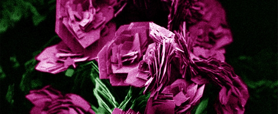
1. BOUQUET OF ROSES
Magnesium hydroxide particles, prepared by a hydrothermal process.
Photographed through a scanning electrons microscopy (SEM).
By: Dana Katz, from the Research Group of Yaron Paz on Photocatalysis and Thin Films, at the Wolfson Department of Chemical Engineering.
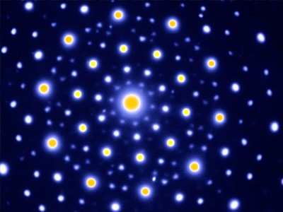
2. GALAXY
Electron diffraction with five-fold rotational symmetry from the icosahedral phase.
Photographed through a transmission electron microscope (TEM).
By: Distinguished Professor Dan Shechtman, from the Department Of Materials Science and Engineering.
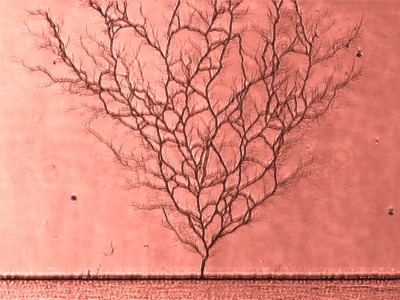
3. FALL
A network of micro and nano-fluidic channels in polydimethylsiloxane (PDMS).
Photographed through an epifluroescence microscope.
By: Merav Karsenty and Nadya Ostromohov, from the Microfluidic Technologies Laboratory headed by Assistant Professor Moran Bercovici, at the Faculty of Mechanical Engineering.
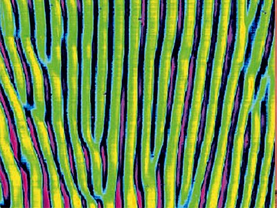
4. FURROWS
Light patterns that form spontaneously when a light beam passes through a nonlinear medium.
Photographed through an optical microscope.
By: The Research Group of Distinguished Professor Mordechai (Moti) Segev, from the Department of Physics.