AI Enables Breakthrough in MRI Efficiency
Researchers at the Technion and in the United States have developed a method combining AI and mathematical models to accelerate and improve medical scans
A group of researchers from the Technion and the United States reports a breakthrough in MRI scanning in a paper published in Nature Communications. The researchers developed an innovative method that accelerates and enhances MRI scans for breast cancer imaging, a disease diagnosed in approximately 2.3 million people each year, most of whom are women.
The new method, called ELITE, combines artificial intelligence with advanced mathematical models, enabling dynamic MRI with unprecedented speed and accuracy. This international study brings together expertise in engineering, MRI physics, artificial intelligence, and clinical radiology.
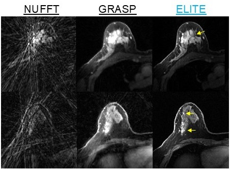
Video and scientific image caption: Demonstration of ELITE results in two patients — a technology that provides high temporal and spatial resolution, reduces noise, and enhances the visualization of breast tumor (marked in yellow) and blood vessels morphology.
Dr. Eddy Solomon of the Technion’s Faculty of Biomedical Engineering, the paper’s lead author, explains that the study focuses on dynamic MRI, a critical technology in breast cancer diagnosis. Dynamic MRI is used primarily for screening populations at high risk for breast cancer and is characterized by exceptionally high sensitivity, with more than 90% accuracy, compared to approximately 50–60% for ultrasound and mammography combined. However, MRI technology faces a major challenge: producing highly detailed images usually requires longer scan times, making it difficult to track the flow of contrast material through the examined tissue. Traditional MRI exams provide one image every 1–2 minutes at best, limiting the ability to accurately capture the fast dynamics of the contrast agent in real time.
Dr. Solomon and his colleagues bridged this gap by combining mathematical modelling that identifies structural and functional patterns in different tissues with a deep neural network (ResNet) trained to remove noise and distortions, along with intelligent reconstruction of missing information from undersampled measurements. The result: generation of one image per second.

The ability to track the movement of the contrast agent almost continuously will allow physicians to identify small tumors more accurately, better distinguish between benign and malignant tumors, and more precisely characterize biological tumor properties such as blood flow and vascular permeability. In a study involving 54 patients, the researchers achieved improved tumor visibility compared to existing methods, exceptionally high image quality, and high diagnostic sensitivity. In addition, shortening scan times is expected to increase the number of women who can be scanned using a given MRI machine.
This study is a direct continuation of a recent research project published a year ago in Radiology: Artificial Intelligence, in which Dr. Solomon and collaborators from New York University (NYU) created a unique repository of 300 breast cancer MRI scans specifically designed for the development of AI-based methods.
Although the method was tested specifically on breast cancer imaging, the researchers demonstrated that ELITE may also be useful for brain, head, and neck imaging. Moreover, the method has the potential to improve not only MRI scans but also other imaging platforms, paving the way for intelligent systems that enable fast, accurate, and personalized imaging while providing physicians with deeper real-time biological insights.
The study included researchers from Weill Cornell Medical College and the NYU Center for Advanced Imaging Innovation and Research. It was supported by grants from the NIH (National Institutes of Health) and RSNA Research (Radiological Society of North America).
Dr. Solomon’s research focuses on developing MRI scanning methods from both the computational and physics perspectives, with the goal not only of accelerating and improving scan accuracy, but also of making MRI technology more accessible to patient populations for whom MRI scanning poses a significant challenge. Additional topics can be found on the research group’s website: https://www.eddysolomonlab.com/
To read the full article, click here