


How does a Protein Recognize its Specific Binding Site on DNA?
Assoc. Prof. Tali Haran unveils the amazing recipes of DNA
DNA can be seen as a cookbook, containing all the recipes needed by the human body. But how is a particular “recipe” picked out? How is it bookmarked? What decides how many portions are cooked? How do some recipes have variants, like a ravioli recipe might have alternative fillings? How are comments written on the margins, such as a good housewife might add: “for green apples, add more sugar”? All these functions are performed on the DNA by regulatory proteins.
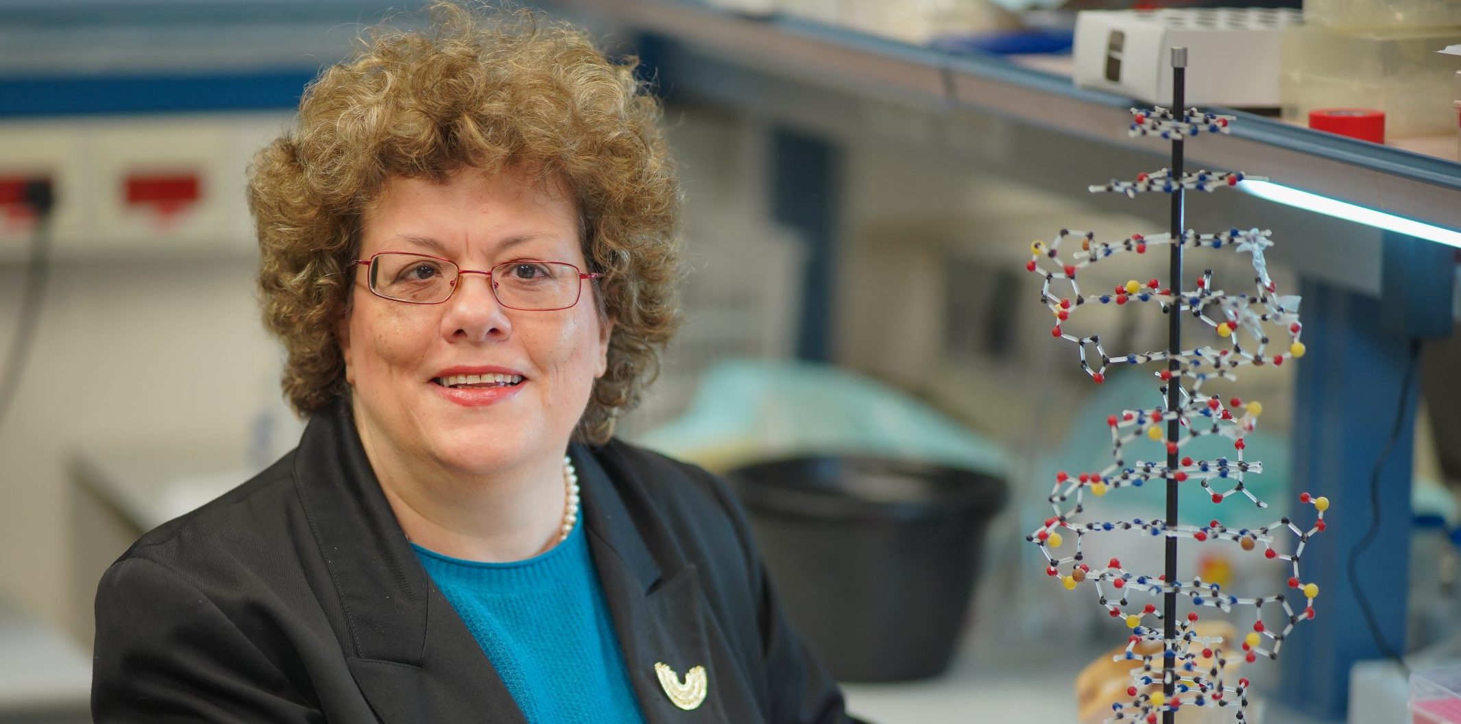
Associate Professor Tali Haran
But how do the regulatory proteins recognize the precise place on the DNA they are supposed to bind? That is the focus of the research of Associate Professor Tali Haran from the Department of Biology at the Technion – Israel Institute of Technology.
In recent years, Prof. Haran’s laboratory has focused on one regulatory protein in particular: p53. Known as the “Guardian of the Genome”, p53 is capable of recognizing multiple types of cellular stress. After doing so, it can then activate DNA repair mechanisms, arrest the cell cycle, or even send the cell into a regulated death process. It thus acts as a tumor suppressor. Indeed, in more than 50% of cancers, p53 is damaged, and its function impaired, so understanding how p53 works is of particular importance.
The functional form of p53 is constructed from four subunits arranged in two pairs. Its activation depends on the binding of the two pairs together while attached to the DNA. There are, therefore, specific binding sites for them on the DNA strands – at each location, there is one binding site composed of two half sites, one for each pair. But there are great variations among these sites. Why this variation? What effect, if any, does it have? This is the question Prof. Haran’s team attempt to answer in their most recent study. (link)
As it turns out, each pair of p53 subunits is semi-attached right from the start – like two cherries joint by a twig. Attaching to their binding site on the DNA brings this pair close together, allowing them to form a more stable attachment. Next comes the joining of the second p53 pair. The DNA is more flexible in some parts, and more rigid in others. (That occurs because of the different building blocks it is made of.) A DNA region that is more flexible can swivel a bit, helping the two p53 subunits within one pair come together and stabilize their attachment to the DNA. A DNA region that is more rigid, by contrast, would not facilitate optimal contacts between the two p53 subunits of one pair and so need the help of the nearby pair residing on a flexible region, or just need more protein to come quickly together to the DNA and form fast a tetramer. An interesting finding of the research is that for most p53 binding sites in the human genome one half site is flexible, whereas the other is more rigid.
The group then proceeded to switch around the two p53 half sites of one specific p53 binding site. They were surprised to find that even such minute changes immediately affected p53 binding. This proves that the differences in the binding sites are not random, but instead allow great precision and nuance in p53’s activity. As for the rigidity/flexibility, this mechanism protects the binding area from being influenced by the sequences flanking the DNA from either side. If for some reason, due to some mutation, the DNA chain is changed and made more rigid or more flexible in the general area where p53 is supposed to get attached to the DNA, the individual binding sites, with their varied rigidity and flexibility, are able to compensate, holding the DNA in just the right shape for everything to come together.
The research team also included Dr. Alon Senitzki, Jessy Safieh, and Dr. Yael Danin-Poleg from the Department of Biology, Technion – Israel Institute of Technology; and Vasundhara Sharma and Professor Alberto Inga from the Department of Cellular, Computational and Integrative Biology (CIBIO), University of Trento.