


Life in Super-Resolution
Researchers from Israel’s Technion have developed a new method for super-resolution single-molecule microscopy with unrivaled efficiency. In a matter of seconds/minutes, it produces images that typically take hours or even days to produce, and could make it possible to visualize nanoscale dynamic processes in real time.
HAIFA, ISRAEL (July 23, 2018) – Researchers at the Technion-Israel Institute of Technology have developed an innovative image reconstruction technique for super-resolution microscopy. The method exhibits unprecedented speed and accuracy, achieving state-of-the-art resolution without any assumptions on the structure in the sample. In a matter of seconds/minutes, it produces images that typically take hours or even days to produce.
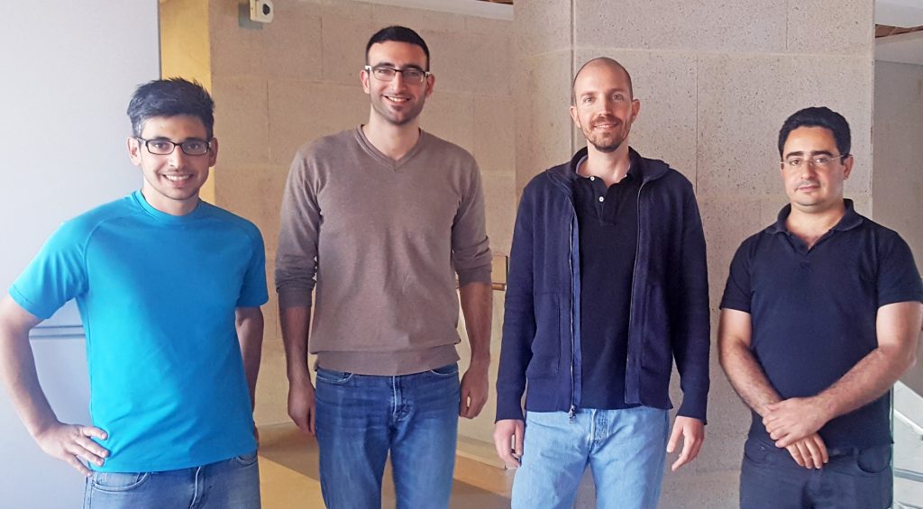
(L to R) Dr. Lucien E. Weiss, Elias Nehme, Assistant Professor Yoav Shechtman and Assistant Professor Tomer Michaeli
The research group was led by Dr. Yoav Shechtman of the Technion Faculty of Biomedical Engineering and Lorry I. Lokey Interdisciplinary Center for Life Sciences & Engineering, and Dr. Tomer Michaeli of the Andrew and Erna Viterbi Faculty of Electrical Engineering. The study, published in Optica, was conducted by student Elias Nehme and post-doctoral researcher Dr. Lucian E. Weiss.
“Typically in super-resolution microscopy, the data is analyzed and an image is produced only after the acquisition is over,” said Dr. Shechtman. “It would be highly desirable to visualize nanoscale dynamic processes in real time, and this technique gets us one step closer to this feat, by enabling ultra-fast image reconstruction.”
In traditional optical microscopes, the resolution (or sharpness) of the image is constrained by the Abbe diffraction limit, a boundary calculated by German physicist Ernst Karl Abbe in 1873. Abbe proved that a microscope’s potential resolution could not go beyond a certain point – approximately half a wavelength. In other words, using wavelengths that are visible to the human eye, it is impossible to distinguish features smaller than 200-300 nanometers.
Of course, the optical microscope has advanced since the 19th century and new methods have succeeded in bypassing the Abbe diffraction limit in order to produce higher resolutions, known as super-resolution imaging. Despite this, the field of nano-biology presents challenges to these new methods, in part because short waves that allow for higher resolutions can harm living cells. Moreover, the dynamic nature of living cells makes imaging speed critical.
Localization microscopy, known as PALM (Photo-Activated Localization Microscopy) and STORM (Stochastic Optical Reconstruction Microscopy), is a recently developed method that produces a single image from a sequence of images (a movie) containing blinking fluorescent molecules. Computerized analysis of this movie, which typically consists of determining the positions of each individual molecule, produces a single super-resolved image with a resolution about 10 times better than the resolution of a conventional microscope.
Even this technology has difficulties reconstructing a single image from the many pictures of flickering molecules. For example, when nearby molecules blink simultaneously, their images overlap and make it difficult to determine their individual positions. Various computational solutions have been designed to solve this problem, but all of them are very complex, have long run times, and demand parameter-tuning, making them difficult to use for the non-experts.
The innovation of the Technion group is to harness the accumulated knowledge in artificial neural networks to aid with the super-resolution imaging problem. Artificial neural networks are a set of algorithms, modeled loosely after the human brain, that are designed to recognize patterns. Their hierarchical structure allows them to analyze complex information and to interpret sensory data through a kind of machine perception, labeling or clustering raw input.
The Technion researchers trained the computer to automatically produce super-resolution images from blinking data, by feeding it images of dense molecules along with their correct positions. The resulting method produces highly accurate super-resolution images directly from the raw fluorescence intensity images, and does so orders of magnitude faster than existing approaches. Moreover, unlike existing methods, this method does not require any parameters, special skills from the user, or previous knowledge about the structure of the sample.
The research was sponsored by Google Research Fund, the Zuckerman Foundation, the Technion – Israel Institute of Technology through its Career Advancement Chairship, the Ollendorf Foundation, the Henry and Marilyn Taub Foundation, the Alon Fellowship program, and the Israel Science Foundation. NVIDIA donated a cutting-edge Titan Xp GPU graphics card to the research team.
Click here for the paper in Nature