


“Science” reports: Technion researchers discover proteins that merge any two animal cells, and even a virus with mammalian cells
 The immediate application is development of innovative technology for treatment of worm parasites
The ability of two or more cells to fuse into one cell is essential for the initial development of an embryo (fusion of sperm and ovum) and for the development of body organs such as the skeleton, muscles and placenta. Despite the importance of the fusion process for human health and reproduction, the cell fusion mechanism is still unknown. Yet, certain enveloped viruses employ similar strategies, which have been deciphered in detail, to fuse and infect body cells of their host (e.g., flu and HIV).
In the laboratory of Professor Benjamin Podbilewicz, of the Technion’s Department of Biology, these processes are being unraveled. Critical proteins mediating the process in animals have now been identified for the first time, and their operating mechanism characterized at the molecular level.
In the research now being published, Ori Avinoam, a doctoral student with Prof. Podbilewicz, discovered that this fusion family (“FF”) of genes, initially discovered by their lab in C. elegans worms, also exists in other organisms and that when transferred to mammalian cells, they are sufficient to force any two cells to fuse. “In the beginning we thought that we had discovered a gene family that exists only in nematodes (worms),” says Prof. Podbilewicz. “We were surprised when we discovered that these genes also exist in other organisms, which makes them, or others similar to them, the leading candidates for being responsible for the fusion process between cells in all kingdoms of life.”
This discovery, in the future, will enable scientists to understand how cells in the human body fuse and then help treat diseases stemming from defects in the fusion process that are liable to cause serious problems in fertility and the musculoskeletal system.
Furthermore, the Technion researchers succeeded in expressing the worm proteins onto the virus-like envelope from Vesicular Stomatitis Virus (“pseudovirus”), which can enter only a single cell, once, but cannot multiply. Normally, these enveloped viruses penetrate cells using proteins located on their envelopes. The Technion researchers switched their natural protein with an FF protein from a worm, and thus engineered a virus-like particle that can infect new types of cells. “In order for the fusion to take place,” explains Avinoam, “this protein must also be on the target cell. Since the nematode parasites have this protein, new pseudo-viruses should recognize and infect them. We can target treatment to each specific parasite.”
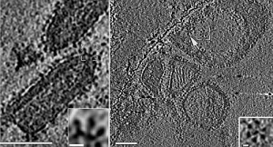
The researchers used electron microscopy and tomography to photograph the protein on the surface of the “pseudovirus” and saw that it creates flower-like structures (see the photograph). Graduate student, Karen Fridman, and researcher, Clari Valansi, also participated in the study, which was conducted in cooperation with Professor Judith White of the University of Virginia in the U.S., Professor Kay Grünewald of Oxford University in England, and Professor Dganit Danino of the Faculty of Biotechnology at the Technion.
Above: The virus-like VSV presenting the AFF-1 protein of the FF family from C. elegans on top of the pseudovirus envelope (left and in the enlargement). Top view (right), shows proteins forming a large complex having the shape of a flower (enlarged – bottom right). The photographs were taken by Dr. Tzvia Zeev Ben-Mordechai from Oxford University and Dr. Ulrike Maurer from the Max-Planck Institute of Biochemistry.